


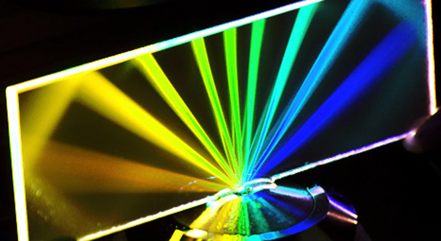
Advanced Light Microscopy
Optical microscopy enables the visualization of structures and biological samples otherwise invisible to the naked eye, providing a magnified image based on their interaction with visible light (absorption, reflection, and refraction). This technique allows not only the characterization of sample morphology and composition, but also the real-time observation of processes occurring at the microscopic scale, such as the diffusion of substances and particles within cells.
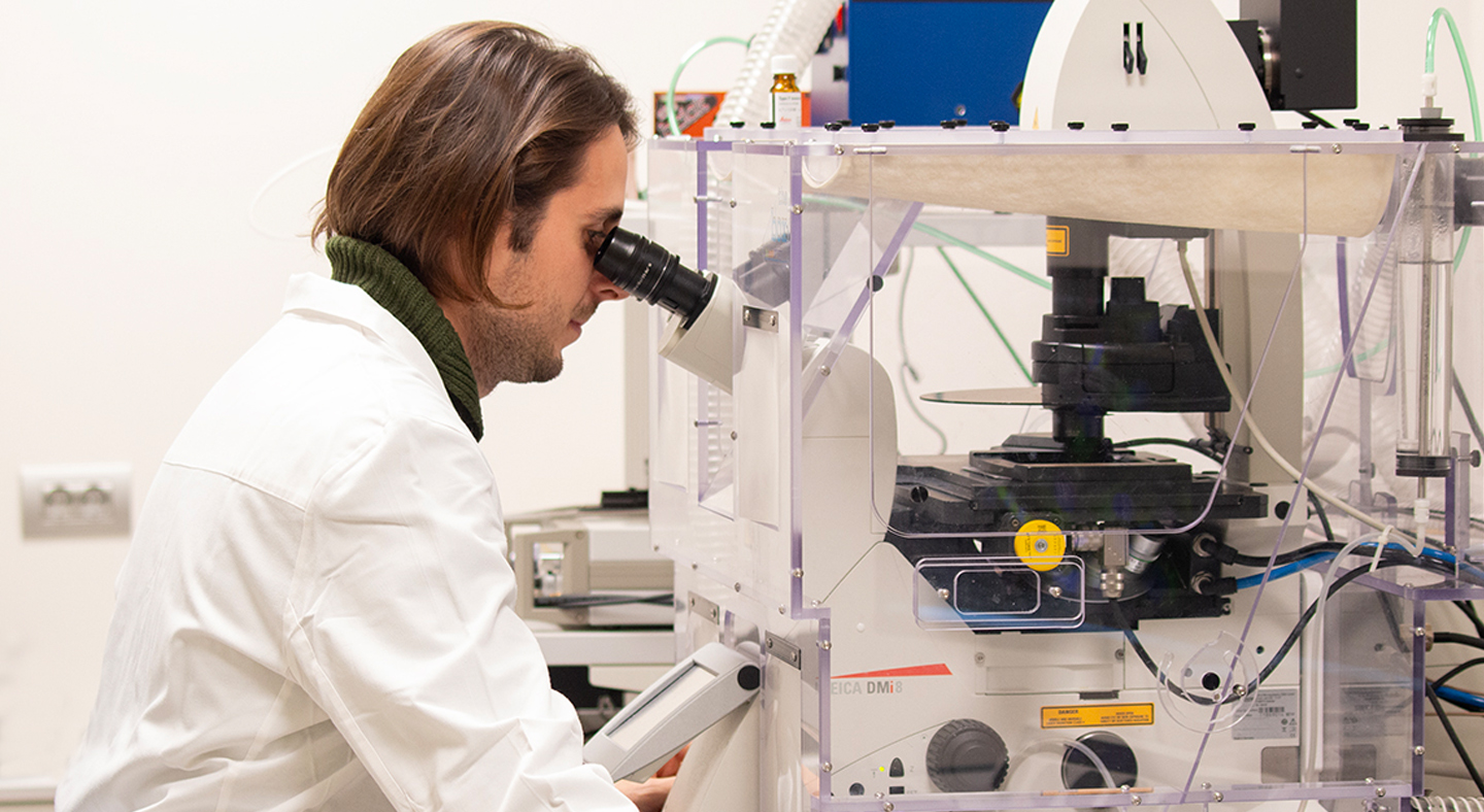
Our unit provides state-of-the-art imaging technologies, including widefield time-lapse microscopy, confocal microscopy, two-photon excitation microscopy, spinning disk confocal microscopy, total internal reflection fluorescence microscopy, and super-resolution microscopy. Our team supports researchers throughout the entire experimental workflow: from experimental design and sample preparation, to image acquisition and analysis, to training in the independent use of instrumentation, while ensuring continuous monitoring of instrument performance to guarantee optimal functioning.
By combining sophisticated imaging technologies with specialized expertise, our goal is to advance the understanding of cancer biology across multiple scales — from single molecules to tissues — thereby contributing to novel scientific discoveries and potential therapeutic developments.
Facility Acknowledgment
According to our Core Facilities Guidelines, if Facility services were used in work described in a publication, the Facility must be acknowledged, citing both the Facillity name and its Research Resource Identifier (RRID). A typically acknowledgement might look like:
The research described was supported by the IFOM Advanced Light Microscopy Core Facility (RRID: SCR_026866)
Co‑Authorship
If a staff member of an IFOM or Cogentech Core Facility contributes substantially to a published work, they should be included as a co‑author.
More information is available in Recommended guidelines and Acknowledging and citing core facilities
Examples of substantial contributions that may justify co‑authorship include:
- Contribution to experimental design or optimization
- Development or significant adaptation of methods or protocols
- Advanced data analysis and interpretation
- Intellectual input influencing the scientific direction of the study
Routine technical support or standard service provision generally warrants acknowledgment rather than co‑authorship.

Head of facility
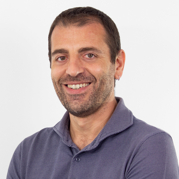
Dario Parazzoli
Dario Parazzoli has built a multidisciplinary background over the years spanning physics, technology, and biomedical research. He graduated in Physics from the University of Milan, where he studied the dispersion of radionuclides following the Chernobyl disaster. He then began his career in the microscopy industry, working for nearly a decade as a project manager and contributing to the development of confocal microscopy and high-end imaging solutions for the life sciences.
Team





Photogallery
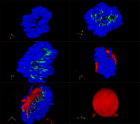
3D rendering of a mitotic He-La cell (blue chromatin, green CREST, red tubulin)
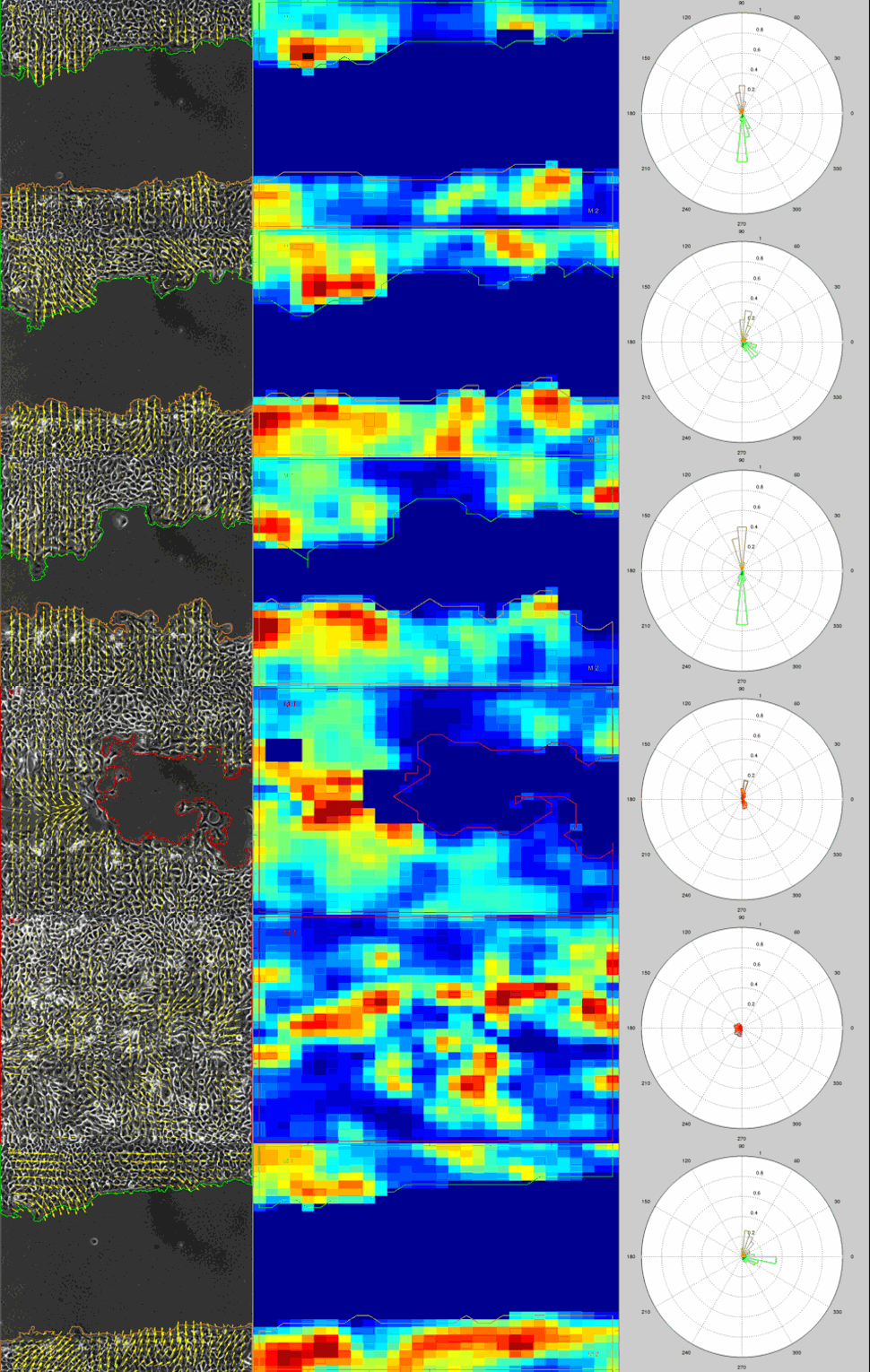
Particle Image Velocimetry (PIV) Analysis of a wound healing experiment
1/2 -