


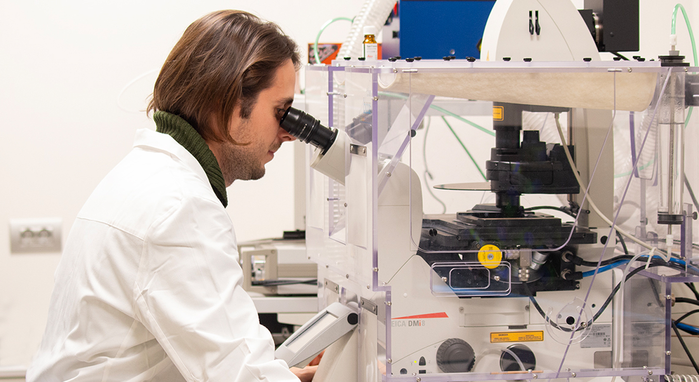
Advanced Light Microscopy
We don't just provide tools: we accompany every project through every phase.
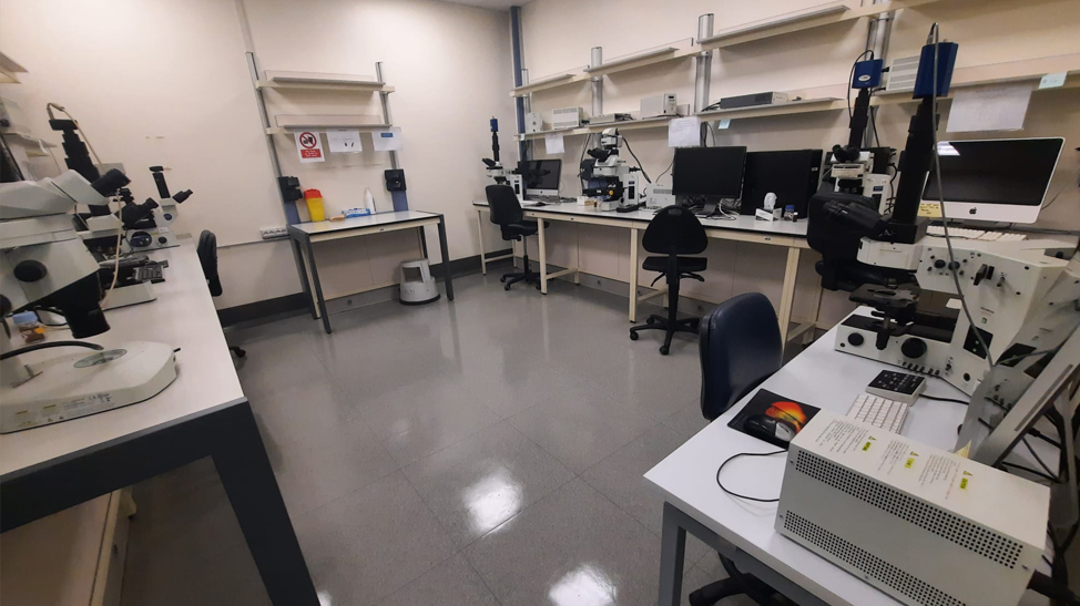
The team of experts supports researchers from sample preparation to data analysis, ensuring a smooth, rigorous, and tailored scientific journey. Whether working with live cells, fixed tissues, or organoids, the facility offers optimized protocols, guided acquisition sessions, and customized analysis pipelines.
Advanced Microscopy Techniques
The page is divided into two parts: imaging platforms, i.e. the instruments used to acquire images, and applications, i.e. the biological questions these platforms can help answer.
Our imaging platforms
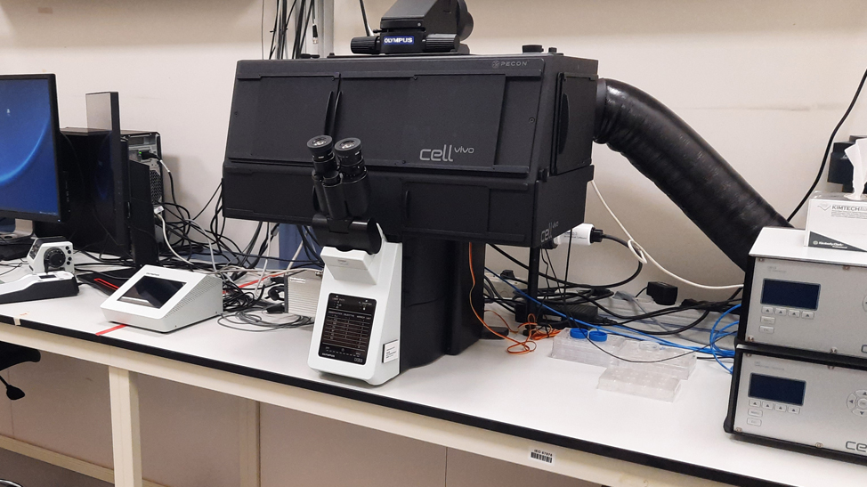
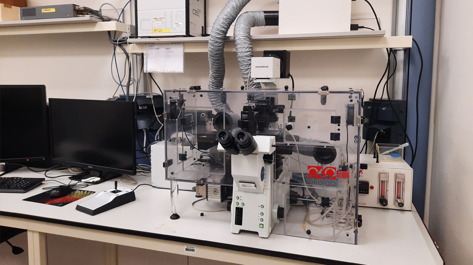
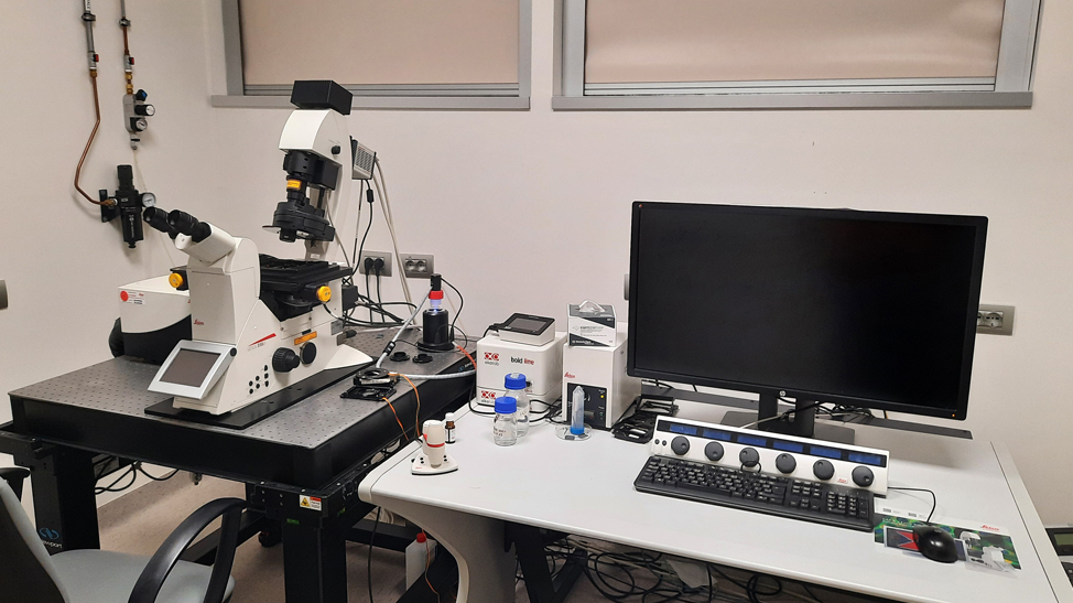
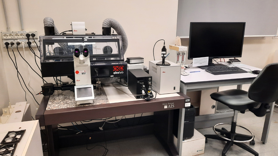
Widefield microscopy
Illuminates the entire sample in a single shot. It is the simplest form of fluorescence microscopy and also the most sensitive, ideal for rapid observations on both fixed and live samples.
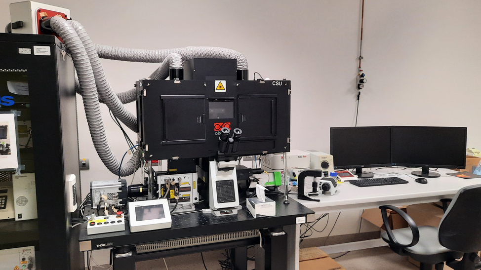
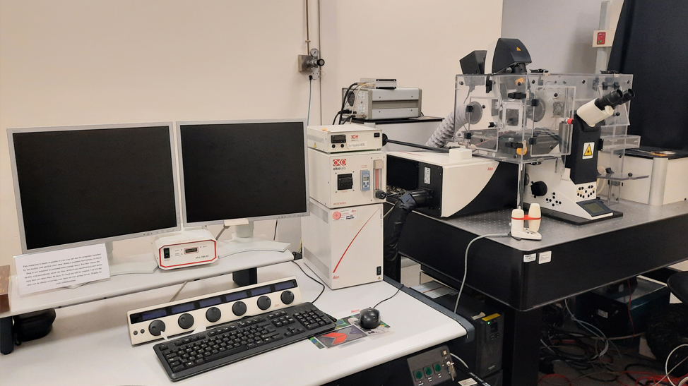
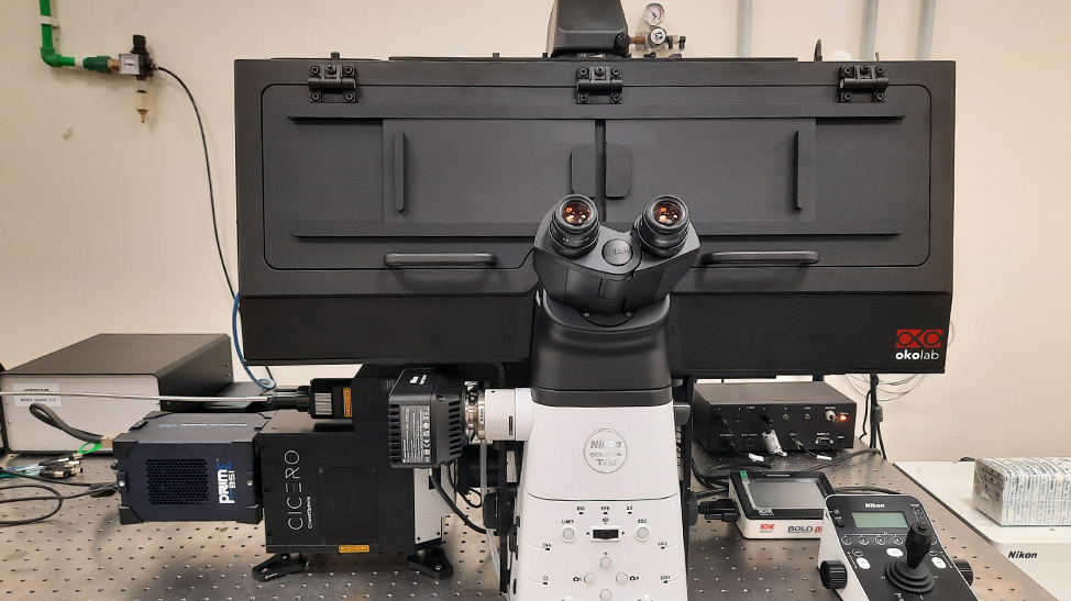
Confocal microscopy
Eliminates out-of-focus light and allows the sample to be reconstructed in three dimensions. It is today the most widely used technique for studying the internal structure of cells and tissues, whether fixed or live.
Light-sheet microscopy
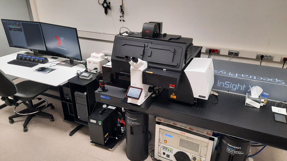
lluminates the sample with a thin "sheet" of light, capturing images very quickly and with minimal damage to cells. Ideal for large, live samples — such as embryos, organoids, and tumor spheroids — observed over long periods of time.
TIRF (Total Internal Reflection Fluorescence)
Illuminates only a very thin region in contact with the coverslip, enabling extremely clean observation of events at the cell membrane, such as adhesion or contacts between cells.
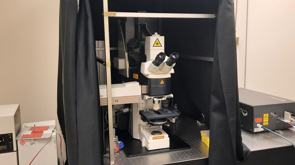
Non-linear microscopy
Two-photon microscopy, second harmonic generation.
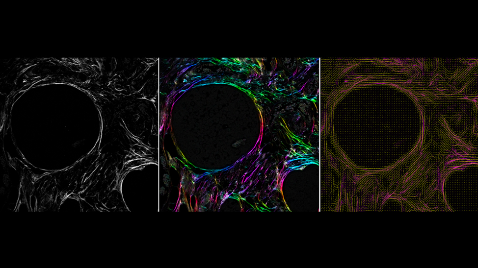
Uses laser light to look deeper into tissues with less damage to cells. Second harmonic generation also makes it possible to visualize structures such as collagen without any staining.
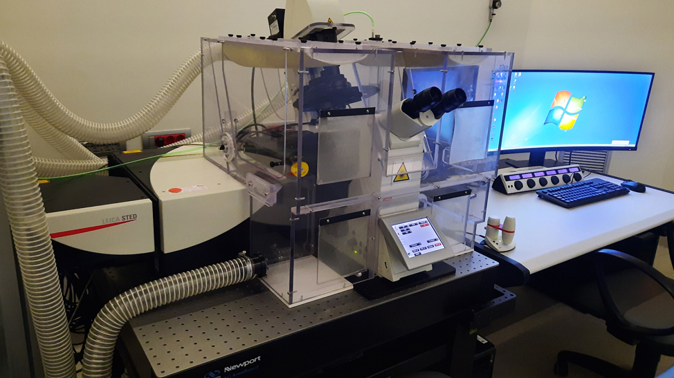
Nanoscopy (super-resolution)
STED.
Surpasses the physical resolution limit of visible light, revealing details up to ten times smaller than those visible with traditional microscopy.
Correlative light-electron microscopy (CLEM)
Combines light microscopy, which shows where a molecule is located and what it does in a living cell, with the ultrastructural resolution of electron microscopy.
Applications
Acquisition on fixed samples and tile scanning
The simplest starting point: a single image of a fixed sample. When the area to be observed exceeds the microscope's field of view, tile/mosaic acquisition automatically stitches multiple fields of view together into one large-format image.
Time-lapse on live cells
The next step is following a living sample over time, to observe processes such as cell division, migration, or changes in shape. Many images are captured over time, using the most suitable platform (widefield, confocal, light-sheet, or others), tracking the process as it unfolds.
Intravital microscopy
Allows direct observation of what happens in a tissue or tumor inside a living organism, in real time. It relies mainly on two-photon microscopy, which achieves imaging depths sufficient to study the behavior of tumor cells in their natural environment, including interactions with the immune system or response to treatment.
Study of molecular movements
Optogenetics, photoactivation, FRAP, FLIP
These methods make it possible to "highlight" a small group of molecules within the cell using light and track their movement over time: specific fluorescent proteins can be selectively switched on, switched off, or displaced, allowing observation of how quickly they diffuse or exchange with neighboring molecules. They are typically performed on confocal or widefield systems. A way to understand how proteins behave — for example, in tumor growth or in response to a drug.
Protein interactions
Colocalization, FRET, fluorescence lifetime imaging
Inside the cell, proteins often work in close contact with one another. These methods, applicable across different platforms, make it possible to verify whether two proteins are truly located at the same point in the cell, or even whether they are close enough to be touching — essential tools for understanding molecular mechanisms that can be altered in cancer.
Laser-induced DNA damage and repair
A laser beam can be aimed at a precise region of the nucleus to create small, controlled DNA damage. By observing what happens immediately afterward, one can watch in real time which proteins intervene to repair the damage — a key aspect in studying the mechanisms that, when altered, can promote tumor development.
Mechanobiology
Cells also respond to physical stimuli, not just chemical ones: how stiff the surrounding tissue is, how much force they receive or exert. These methods make it possible to measure, and in some cases reproduce, these mechanical forces, which are important in tumor growth and spread.
Microinjection
Using precision instruments, substances — DNA, proteins, dyes — can be injected directly into a single living cell, useful for testing the effect of a molecule in a targeted way.
To determine which platform or method is best suited to their research project, researchers can contact the Advanced Light Microscopy Unit: our team is available to help define the most appropriate experimental approach.
Sample Preparation Support
In microscopy, sample preparation is a fundamental step for obtaining high-quality images and reliable data. Preparation methods vary depending on the type of sample, the acquisition techniques used, and the objectives of the analysis.
We support researchers in this phase, advising on the most suitable protocols for different experimental needs, and we assist them in experimental design, for example in the choice of specific reagents such as fluorescent proteins, sensors, or dyes.
We also provide reagents useful for staining fixed samples (secondary antibodies labeled with fluorophores, nuclear dyes...) and for sample mounting (mounting medium).
Image Analysis & Data Processing
With approximately 180 custom analysis protocols developed annually, our team provides support immediately after the collection of the data, from image processing to data analysis and interpretation. All the plugins and pipelines can be developed in open-source software (Fiji, CellProfiler, QuPath) or commercial platforms (Matlab, Arivis, Harmony, Prism). Examples of implemented tools include:
Cell, Nuclei, and Particle Detection:
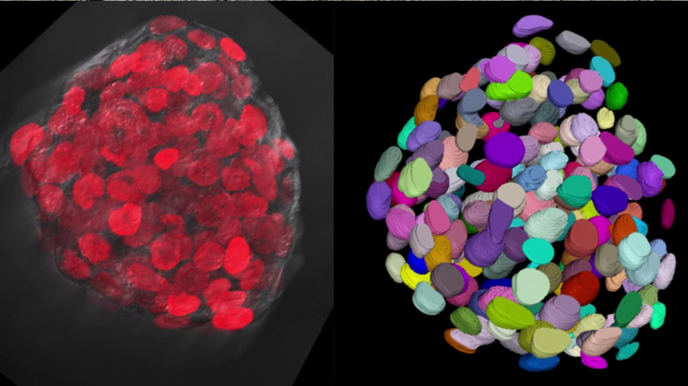
Segmentation and classification using classical methods or machine learning tools (2D and 3D).
Cell and Particle Tracking:
Pipelines for single-cell tracking, Particle Image Velocimetry (PIV), Wound Healing Assays, and particle trafficking.
Molecular Mobility:
FRAP analysis.
Label-free imaging analysis:
Machine learning tools for collagen fiber orientation analysis.
Protein Interactions:
Quantification of colocalization, foci formation (2D and 3D), and FRET analysis.
Immunohistochemistry imaging analysis:
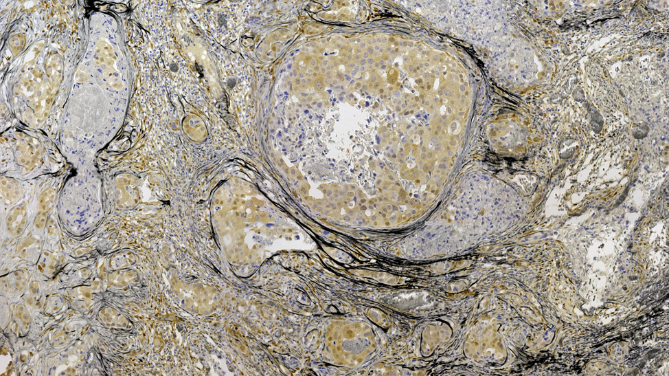
Object detection pipelines using color threshold segmentation or machine learning (pre-trained or custom-trained models), applicable to whole-slide and single-image analyses.
AI-Assisted Image Analysis:
Integration of artificial intelligence and machine learning tools for enhanced segmentation, tracking, and quantitative microscopy workflows.
Training & Support
We provide comprehensive support tailored to both senior researchers and early-career scientists, offering personalized training to foster their professional development. Our team ensures that users gain the necessary expertise to fully exploit the advanced microscopy resources available at IFOM.
1/3 -
Microscopy Applications
Seconda armonica: organizzazione del collagene in una sezione istologica di carcinoma mammario